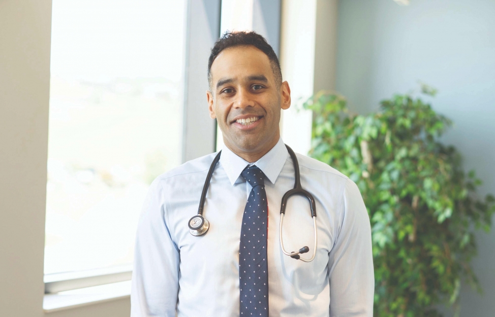
My role
I am a cardiologist at Monument Heart and Vascular Institute with a primary focus in cardiovascular imaging. I grew up in Rapid City and did my medical and cardiovascular training at New York Presbyterian. Additionally, I did a year of advanced cardiovascular imaging with a focus on cardiac CT, structural echocardiography and cardiovascular MRI at Houston Methodist Hospital. I help manage cardiovascular patients at Monument Health in the clinic and the hospital as well as assist in the specialized imaging needed for the Monument Health Heart Failure team as well as the Monument Health Structural Heart team.
My goal
My goal is to provide the Rapid City community and our patients with the newest technological advancements in cardiovascular care and cardiovascular imaging. I strive
to continue to learn and evolve as a physician and a
cardiologist so that I can provide our community with the most appropriate and optimal care. I hope to help the cardiology practice in Rapid City evolve and grow so that patients can receive their care close to home.
My passion
My passion is serving people in their greatest time of need. Cardiology provides an opportunity to help people in their most vulnerable moments in the hospital and then help and support them as they work to improve their heart health once they go back home. I am eager to contribute my skills and dedication to make a difference in the lives of those facing cardiovascular challenges, and feel fortunate that I am able to do so.
Dr. Ameer focuses on advanced cardiac imaging. This specialty refers to sophisticated techniques and technologies used to obtain detailed and comprehensive images of the heart and surrounding structures. This includes:
- Cardiac magnetic resonance imaging, which provides detailed images of the heart anatomy and function using
- a strong magnetic field and radio waves.
- Cardiac computed tomography, which uses X-rays to create cross-sectional images of the heart, allowing for detailed visualization of the coronary arteries and other structures.
- Echocardiography, which uses sound waves to create images of the heart and provides real time information about structure and function.
- Nuclear cardiology, in which a small amount of radioactive material is injected and used to assess blood flow and function of the heart muscle.